


Nanoparticles are frequently synthesised as colloids, dispersed in solvents such as water, hexane or ethanol. For their characterisation by transmission electron microscopy (TEM), a drop of colloid is typically deposited on a carbon support and the solvent allowed to evaporate. However, this method of supporting the nanoparticles reduces the visibility of fine atomic details, particularly for carbonaceous species, due to interference from the 2-dimensional carbon support at most viewing angles. We propose here the impregnation of a 3 dimensional carbon black matrix that has been previously deposited on a carbon film as an alternative means of supporting colloidal nanoparticles, and show examples of the application of this method to advanced TEM techniques in the analysis of monometallic, core@shell and hybrid nanoparticles with carbon-based shells.
Nanoparticles represent one of the most studied structures in nanotechnology and nanoscience because of the wide range of applications arising from their unique optical, physical and chemical properties [1]. Often they have core@shell structures, or are coated with organic molecules. Nanoparticle functionality is largely affected by the specific configuration of the outer surface atoms. For example, in heterogeneous catalysis activity and selectivity are mostly determined by the type of atomic defects present at the surface of metallic nanoparticles, and in the field of biomedicine the surface coating of hybrid (inorganic core@organic shell) nanoparticles regulates their stability, solubility and targeting.
Nanoparticles are frequently synthesised using solution techniques that yield colloids, i.e., a solid–liquid mixture containing solid particles that are dispersed to various degrees in a liquid medium; most frequently water, ethanol or hexane. Colloid characterisation generally employs a variety of techniques to establish understanding and control over nanoparticle synthesis and properties. Electron microscopy in transmission mode (TEM) and in scanning transmission mode (STEM) are widely used for particle characterisation, and advances in these techniques mean that it is now routinely possible to resolve single atoms at the surfaces of nanoparticles using aberration-corrected microscopes, to elucidate the three-dimensional shapes of nanoparticles using electron tomography, and to enhance the contrast in very low density materials (e.g., carbonaceous materials) using electron holography [2] and [3]. However, the significant potential of these (S)TEM techniques is ultimately limited by the sample and the techniques available for sample preparation.
Typically, examination by (S)TEM requires that a nanoparticulate sample be prepared by depositing a drop of colloid on a thin, electron-transparent support. It is usual that an amorphous carbon film, silicon nitride film or graphene layers deposited on a copper grid constitute the support [4]. Crucially, these sample preparation techniques suffer from the major limitation that the contrast from the support often shadows atomic details at the particle surface. Moreover, it has been established that the thinnest supports can degrade under electron-beam irradiation, affecting particle stability [5], and also that hydrocarbon contamination can be an issue [6]. The most widely used commercially available TEM support is holey carbon, which comprises of a perforated carbon thin film. In this case, sample preparation aims to locate at least some of the nanoparticles of interest at the edges of the perforations. However, the concave nature of the holes means that solvent contaminants tend to accumulate preferentially at these sites. Moreover, if the TEM sample holder is tilted a particle attached to the edge of a hole is very likely to be shadowed by the carbon film. Taken together, these drawbacks significantly limit the application of techniques such as electron tomography [6].
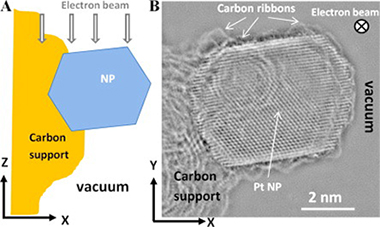
We propose here a method of circumventing some of these fundamental problems by developing a technique for mounting nanoparticulate samples using a carbon matrix that is inspired by the way samples used in electrocatalysis are prepared [7]. Fig. 1 shows an image of a typical Pt-based electrocatalyst supported on carbon black as used in proton-electron membrane fuels cells, and which consists of Pt nanoparticles formed by calcination of a carbon black impregnated with a solution of salt precursor. Carbon black is a low-grade form of graphite, which is composed of nanocrystallites and no long-range order [8]. In Fig. 1 the carbon black is Vulcan XC-72R, which is widely used as a catalyst support in fuel cells because it provides high electrical conductivity, good reactant gas access, adequate water handling and good corrosion resistance, whilst allowing high dispersion of the particles. In electrocatalyst samples it is common to find particles, like the 5 nm Pt particle shown in Fig. 1, attached strongly to the surface of the support and viewed edge-on against a vacuum so as to provide optimal conditions for high-resolution TEM (HRTEM). Fig. 1B is a quantitative phase image of a Pt particle obtained from a defocus series of 20 images at intervals of 5 nm acquired in a FEGTEM JEOL 2020 at 200 kV with spherical aberration of −30 μm and applying the exit-wave restoration technique [2]. The contrast between details of the particle finestructure is very high compared to conventional HRTEM images, and details such as the presence of monoatomic carbon ribbons surrounding the particle can be seen.